

AMD Treatment & Prognosis

Ophthalmologist, Retinal Specialist
Pacific Eye Institute
February is Age-Related Macular Degeneration (AMD) month. A deterioration of the retina and choroid that leads to a substantial loss in visual acuity, AMD is the leading cause of significant visual acuity loss in people over age 50 in developed countries.
At Pacific Eye Institute, we make an AMD diagnosis by a clinical examination with a slit lamp and by using several types of imaging including fluorescein angiography (FA) and optical coherence tomography (OCT).
Treatment and Prognosis
Dry AMD: Based on a study conducted by the National Eye Institute, the AREDs-2 nutritional supplement formula may delay and prevent intermediate dry AMD from moving to the advanced form. The AREDS supplement formula, which is widely available over the counter, contains:
- Vitamin C
- Lutein
- Vitamin E
- Zeaxanthin
Wet-AMD treatment has been revolutionized in recent years after the discovery of vascular endothelial growth factor (VEGF), a family of compounds in the body. There are currently 4 anti-VEGF drugs:
- Avastin® (bevacizumab)
- Lucentis® (ranibizumab)
- Eylea® (aflibercept)
- Beovu (brolucizamab)
3 Anti-VEGF Treatment Regimens
- Pro re nata (PRN) or “treat and observe:” patients are treated with 3 initial monthly injections followed by treatment as needed
- “Treat and extend:” after 3 initial monthly injections, the time between treatments is gradually increased until wet AMD is stabilized
- Monthly injections
Everyone is different but the approach I like to take is “treat and extend.” Patients tend to have similar outcomes and will have decreased treatment needs, making compliance and patient satisfaction higher while ensuring good vision.
More Information About AMD
Demographics: As many as 11 million people in the United States have some form of age-related macular degeneration (AMD). This number is expected to double to nearly 22 million by 2050. The number of people living with macular degeneration is expected to reach 196 million worldwide by 2020 and increase to 288 million by 2040.
Causes: The condition develops as the eye ages. Some have been linked with higher risk (see below).
Types: There are 2 types of AMD: non-neovascular or dry AMD; and neovascular or wet AMD.
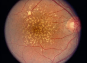
Dry AMD: In early stages of dry AMD, the hallmark is drusen—pale yellow lesions formed beneath the retina. Drusen are made up of lipids and as they accumulate, dry AMD can progress. (Photo 1)
Photo 1: Patient with dry intermediate drusen
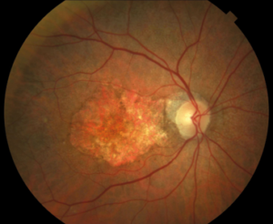
Atrophic areas in the RPE also may develop; if the atrophic area is significant and with sharp borders, it is termed geographic atrophy (GA). GA is the advanced form of dry AMD which is frequently associated with loss of central vision. (Photo 2)
Photo 2: Patient with geographic atrophy
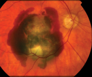
Wet AMD: In wet AMD, there is a sudden or gradual decrease in visual acuity, blind spots in the center of vision and distortion of straight lines. The hallmark of wet AMD is choroidal neovascularization (CNV). CNV occurs when abnormal blood vessels grow beneath the retina; these can bleed (Photo 3) or leak and cause a distortion.
Photo 3: Patient with large CNVM with subretinal hemorrhage
Risk Factors
- Age—the strongest risk factor
- Family history of AMD
- Caucasian race
- Cigarette smoking
Other Possible Risk Factors
- Female gender
- High cholesterol
- Lower level of education
- Sunlight exposure
- Light iris color
- Low dietary fish intake
- Farsightedness
- Higher body mass index (BMI)
- Cardiovascular disease